Aging has been always a significant concern for mankind and they have been seeking a solution to overcome this challenge from the beginning. Biology as a scientific method presents important approaches which can revolutionize aging studies. Approaches that improve understanding of the underlying molecular mechanisms of aging, as well as their contributions to age-associated diseases. Studying the replicative aging phenomenon in the budding yeast has led to significant findings on how aging is regulated by evolutionarily conserved enzymes and molecular pathways. Identifying and characterizing the factors that modulate longevity is central to understanding the basic mechanisms of aging. Among model organisms used for research related to aging, the budding yeast has proven to be an important system for defining pathways that influence lifespan. Replicative lifespan is defined by the number of daughter cells a mother cell can produce before senescing. Over the past 10 years, replicative life span analysis has been performed on several thousand yeast strains, identifying several hundred genes that influence replicative longevity.1
Based on the conservative method of aging studies, tracking of mother cells and observing molecular markers during the process of aging face different limitations. Fifty years after Mortimer and Johnston’s discovery, the technology used to analyze replicative aging remained essentially the same. To measure the number of daughter cells produced by each mother cell, Mortimer and Johnston grew yeast cells on an agar plate and used a micromanipulator (a microscope with a dissector) to remove daughter cells after each cell division2. This is still the most widely used method for analyzing yeast lifespan. However, because the cells are grown on an agar plate, it is almost impossible to follow cell and organelle morphologies and track molecular markers throughout the lifespan of individual cells. Such high-resolution, single-cell analysis is critical for developing a mechanistic understanding of cellular aging and death. In addition, the traditional assay is laborious and time-consuming, which makes it very difficult to perform large-scale screening for mutants with lifespan phenotypes.3
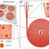
Recently, the microfluidic system which is capable of retaining mother cells in the micro pads while removing daughter cells automatically, making it possible to observe fluorescent reporters in single cells throughout their lifespan. Figure 1 shows a microfluidic device for monitoring the aging process of budding yeast.

Figure 1. (a) Array of micropads. The chip contains 200 micropads arranged in an array format(b) Schematic illustration of microfluidic dissection. Mother yeast cells are held between a soft PDMS pad and thin cover glass.4 Copyright (2018) National Academy of Sciences, U.S.A.
The basic unit of the device consists of a microfluidic device with micro pads that can physically trap the mother cells while allowing the removal of daughter cells (Video 1). There are two significant mechanisms for the separation of mother and daughter cells from each other in microfluidics. The first one is separation based on the cells sizes which means that mother cells are trapped in micro pads duo to their bigger sizes and daughter cells are removed by fluid flow. However, as the mother cells get older their and their daughter sizes get bigger that this phenomenon can disrupt the procedure. Geometric confinement by itself alone is not good enough solution because it is sensitive to the height of the micro pad: if it is too high, the mother cells will not be stably trapped; if it is low enough to stably trap mother cells, there is a certain probability that daughter cells will be trapped and jam the device. Modifying the mother cell surface to create adhesion between micro pad walls and Mother cells surfaces is another way for separation. In this method, mother cells are trapped by a combination of geometric confinement (the height of the micro pad is comparable to the size of mother cells) and adhesion between biotin labeled mother cell surface and BSA-Avidin modified glass.
Video 1.4 Copyright (2018) National Academy of Sciences, U.S.A.
Challenges:
However, many issues prevent the use of microfluidic devices in a high-throughput manner for lifespan screens.5
- First, although the time required to monitor the entire lifespan of the yeast cell has been dramatically reduced, the throughput is limited to 1-4 channels per device.
- Second, the basis of this dissection method is the size of cells but the point is that as the mother cells get older, they generate large daughter cells that also become trapped by the micro pads.
- Third, the micro pad design often allows more than one cell to be trapped; multiple cells can be trapped underneath one micro pad, whereas no cells are trapped under others.
- Finally, in some cases, cell-surface labeling and chemical modification of the device are required, which has proven to be technically challenging for fabrication and to introduce adverse effects on replicative lifespan.
Microfluidic dissection platform that allows us to track single yeast cells over their entire replicative lifespans with high-resolution imaging capability, is a possibility that the yeast-aging research community has long-awaited for it. Not only can this technology replace the tedious manual microdissection methods to determine lifespan data, but it can also be used for high-resolution in vivo fluorescence imaging of aging cells under exactly controlled environmental conditions6. This technology thus opens up unique possibilities for future aging research:Its capability for phenotypic tracing is essential (i) to explore the relevance of cell-to-cell heterogeneity in the aging process, and (ii) to address important questions, such as whether certain phenotypes in cellular youth affect old-age behaviour, or how damage is asymmetrically inherited by the mother cell and removed from the daughter cell, and how this changes with age. By elimination of mentioned disadvantages, there are promising signs that this technology will enable novel investigations in the quest for molecular mechanisms underlying the aging process and will permit large-scale screens into the aging phenotype for its capability to be easily multiplexed and to allow aging experiments in an unsupervised manner.
1. Replicative life span analysis in budding yeast. GL, Sutphin, JR, Delaney and M, Kaeberlein. 2014, Methods in molecular biology, Vol. 1205, pp. 341-357.
2. Single Cell Analysis of Yeast Replicative Aging Using a New Generation of Microfluidic Device. Zhang, Yi , et al. 11, 2012, PLOS ONE, Vol. 7, pp. 1-10.
3. Molecular phenotyping of aging in single yeast cells using a novel microfluidic device. Xie, Zhengwei, et al. 2012, Aging Cell, Vol. 11, pp. 599–606.
4. Whole lifespan microscopic observation of budding yeast aging through a microfluidic dissection platform. Lee, Sung Sik , et al. 13, 2012, PNAS, Vol. 109, pp. 4916–4920.
5. High-throughput analysis of yeast replicative aging using a microfluidic system. Jo, Myeong Chan , et al. 2015, PNAS, Vol. 112, pp. 9364–9369.
6. A simple microfluidic platform to study age-dependent protein abundance and localization changes in Saccharomyces cerevisiae. Cabrera, Margarita, et al. 5, 2017, Microbial Cell, Vol. 4, pp. 169-174.
Enjoyed this article? Don’t forget to share.

Mohammad Ali Zoljalali
Mohammad Ali Zoljalali is a Master's degree student at the University of Mazandaran. He is working on microfluidics by the focus on microchannels heat sinks and Drug Delivery.


