Cells form the basic unit of life and can broadly vary in biological structure and function. Nucleic acids encode the information of life by programming cellular functions at the level of transcription for specific biological outcomes. Single-cell genomics provides a window to characterize the identity and function of cells. Drawbacks of the technique include a lack of practical ease and scale. Sequencing information provides a map to understand the properties of a single cell; including the type, state and nature of its biology during its regular function and during pathogenesis. Since all cells are not alike, it is increasingly important to recognize the difference between cells for deeper insight into their behavior for tailored treatments in personalized medicine. It is possible to probe the properties of individual cells in the lab using advanced single-cell sequencing techniques.
The process can be performed primarily using mRNA extracts of cells to determine the level of gene expression. For effective RNA-sequencing at the level of the single cell, life scientists must compartmentalize cells in a volume of comparable size to the cell itself. This process can be accomplished in the lab using a microfluidic device. Nevertheless, an existing demand remains to establish a simpler practical method to extend sequencing to a greater number of cells in parallel, while providing single-cell resolution. Preceding methods for cell profiling were only applicable to hundreds or a few thousand cells. As a result, scientists have developed droplet microfluidics as a fundamental method to meet the demand for fast and scalable approaches.
In droplet-based single-cell sequencing, individual cells are compartmentalized in nanolitre-scale aqueous environments that act as tiny reaction chambers for PCR and for reverse transcription. The method can analyze thousands of cells in parallel for cellular mRNA transcripts while recording the transcript’s cell of origin using a genomic barcodes strategy. The barcodes provide a molecular memory, acting as an identifier- read at sequencing to decode specific information of each individual cell. Two recent papers by A. Klein et al. and E. Macosko et al. introduce the technique of droplet microfluidics for single-cell RNA-sequencing to establish the basic concepts (Video 1). Several commercial kits have rapidly followed the protocols and similar publications, to provide easy access to the technique in the lab.
![]()
Video 1: Trimmed video abstract of microfluidics-based nanoliter droplet formation for highly parallel genome-wide expression profiling of individual cells. Credit: Cell, DOI: https://doi.org/10.1016/j.cell.2015.05.002
The field is advancing rapidly, with many studies aiming to gather data and explore cellular information accessed with the technique. Microfluidics platforms have advanced to integrate within lab-on-a-chip devices to streamline the process at the nanoscale and dissect transient transcriptional states at the level of the single-nucleus. To achieve higher accuracy in the method, scientists have created droplet libraries that can correct composition bias and sequencing errors affecting cellular and molecular barcodes. The sequencing methods can be extended to probe information beyond mRNA as well. For instance, Guo et al. introduced ultra-high throughput, PCR-free, single-cell miRNA assays in a continuous flow microfluidic process. Droplet-based microfluidics has in this way originated major advances in the field at the interface between technological advancements and biomedical applications of single-cell sequencing.
Fundamental steps of the droplet-based single-cell sequencing method can be summarized as follows:
- The process starts with the preparation of a single-cell suspension from a tissue of interest in the lab.
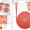
- Scientists then prepare the barcoded primers as surface attachments on simple microparticles or embedded within hydrogel microparticles.
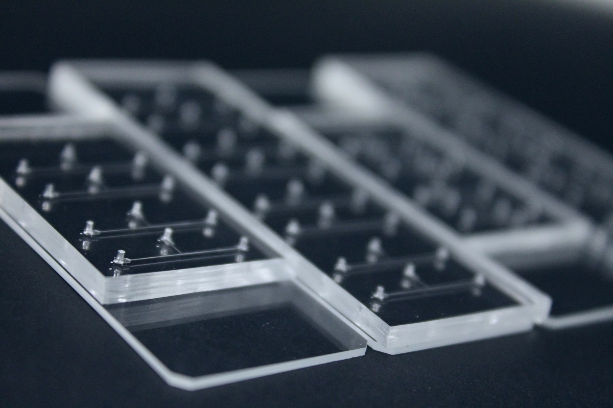
- A microfluidic device is used to encapsulate individual cells in parallel with a distinctly barcoded microparticle within a tiny droplet.
- After the cells are isolated in droplets, the cells are lysed to release the mRNAs, which hybridize to the primers.
- The scientists break the droplets to generate single-cell transcriptomes attached to the microparticles (STAMPs).
- They amplify the STAMPs with PCR, sequence and analyze the transcript’s cell of origin using the STAMP barcodes.
Two types of beads (microparticles); known as simple and hydrogel can be used to synthesize primers. Each microparticle contains a sequence with a PCR handle, cell barcode, a unique molecular identifier (UMI) and a 30-base pair oligodT sequence at the end of all primer sequences. After preparing the single-cell suspension and the microparticles, the individual cells can be encapsulated together with microparticles in droplets, using a custom-designed intricate microfluidic device. Recent work by life scientists in the McCarroll lab detail the individual steps to synthesize cell-barcodes and UMIs in drop-sequencing, where UMIs act as unique identifiers to prevent double-count sequence reads.
As ongoing high-throughput genome sequencing techniques become practically viable, scientists are beginning to use software tools to visualize the expression of data sets. The functions include routine assembly, annotation and display of genomic information as physical maps. For instance, Lohse et al. describe a suite of tools to generate physical maps of plastid and mitochondrial genomes, known as “Organellar Genome Draw”. The freely available web tool with recent upgrades can mainly analyze large-scale, plant-cell derived nucleic acid and protein sequences. In this way, high-throughput, droplet-based single-cell sequencing is rapidly advancing with technical upgrades and accompanying software to broaden scientific understanding of genomics research at the level of the single cell and single nucleus.
Enjoyed this article? Don’t forget to share.

Thamarasee Jeewandara
Thamarasee Jeewandara is an Early Career Scientist and Science Writer, with a Ph.D. in Medicine and Bioengineering from the Sydney Medical School, University of Sydney, Australia. She is currently living in New York, focusing on her research and writing interests in materials science, tissue engineering, and bone research. Find her on Twitter @Jeew333T and her science writing portfolio on Nature Research Bioengineering.