11 Jun Microfluidic co-culture facilitates cross-talk between lung cancer cells and brain perivascular tumor microenvironment
Abstract
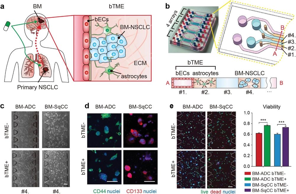
“Non-small cell lung carcinoma (NSCLC), which affects the brain, is fatal and resistant to anti-cancer therapies. Despite innate, distinct characteristics of the brain from other organs, the underlying delicate crosstalk between brain metastatic NSCLC (BM-NSCLC) cells and brain tumor microenvironment (bTME) associated with tumor evolution remains elusive. Here, a novel 3D microfluidic tri-culture platform is proposed for recapitulating positive feedback from BM-NSCLC and astrocytes and brain-specific endothelial cells, two major players in bTME. Advanced imaging and quantitative functional assessment of the 3D tri-culture model enable real-time live imaging of cell viability and separate analyses of genomic/molecular/secretome from each subset. Susceptibility of multiple patient-derived BM-NSCLCs to representative targeted agents is altered and secretion of serpin E1, interleukin-8, and secreted phosphoprotein 1, which are associated with tumor aggressiveness and poor clinical outcome, is increased in tri-culture. Notably, multiple signaling pathways involved in inflammatory responses, nuclear factor kappa-light-chain-enhancer of activated B cells, and cancer metastasis are activated in BM-NSCLC through interaction with two bTME cell types. This novel platform offers a tool to elucidate potential molecular targets and for effective anti-cancer therapy targeting the crosstalk between metastatic cancer cells and adjacent components of bTME.”

“Schematic illustration of brain metastatic niche of NSCLC cells and microfluidic device for recapitulating the niche. a) The brain metastatic niche involving bTME with bECs and astrocytes. b) Representative photography and drawing of the microfluidic device with seven channels, and its cross-sectional illustration with three types of cells. c) Phase-contrast images of tumor cells monocultured (bTME-) and co-cultured (bTME+) in a microfluidic device. d) Representative fluorescence images of patient tumor cells of cancer stemness markers. e) Representative fluorescence images of patient tumor cells tagged by live/dead kit (left), and measured viability (right). Scale bars indicate 200 µm. The viability data presented as average values ± SEM, n = 65 for BM-ADC and n = 21 for BM-SqCC.” Reproduced under Creative Commons Attribution 4.0 International License from Kim, H., et al., Recapitulated Crosstalk between Cerebral Metastatic Lung Cancer Cells and Brain Perivascular Tumor Microenvironment in a Microfluidic Co-Culture Chip. Adv. Sci. 2022, 2201785.
Figures and the abstract are reproduced from Kim, H., Sa, J. K., Kim, J., Cho, H. J., Oh, H. J., Choi, D.-H., Kang, S.-H., Jeong, D. E., Nam, D.-H., Lee, H., Lee, H. W., Chung, S., Recapitulated Crosstalk between Cerebral Metastatic Lung Cancer Cells and Brain Perivascular Tumor Microenvironment in a Microfluidic Co-Culture Chip. Adv. Sci. 2022, 2201785. https://doi.org/10.1002/advs.202201785 under Creative Commons Attribution 4.0 International License.
Read the original article: Recapitulated Crosstalk between Cerebral Metastatic Lung Cancer Cells and Brain Perivascular Tumor Microenvironment in a Microfluidic Co-Culture Chip


