30 Jan Unlocking the Secrets of Bacterial Motility in Porous Microstructures: A Microfluidic Study
Bacteria have a crucial survival strategy of motility which helps them navigate in porous environments. These environments are essential for biogeochemical processes in quiescent wetlands and sediments. However, the understanding of self-transport mechanisms in confined interstices of porous media is limited. In this week’s research highlight, a Communications Physics paper sheds light on the movement of bacteria (Magnetococcus marinus) in a microfluidic chip mimicking the porous media with diverse geometries made using microfabrication techniques. The microfluidic device precisely tracks the cells’ motion and helps understand the interactions between cells and solid matrix surfaces.
“Here, we precisely track the movement of bacteria (Magnetococcus marinus) through a series of microfluidic porous media with broadly varying geometries and show how successive scattering events from solid surfaces decorrelate cell motion.“, the authors explained.

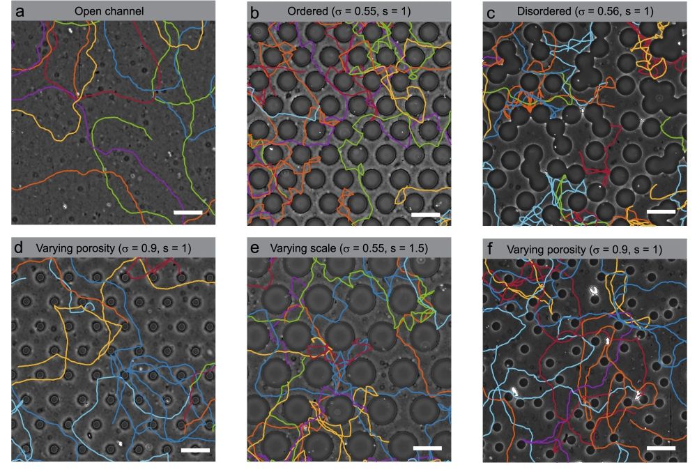
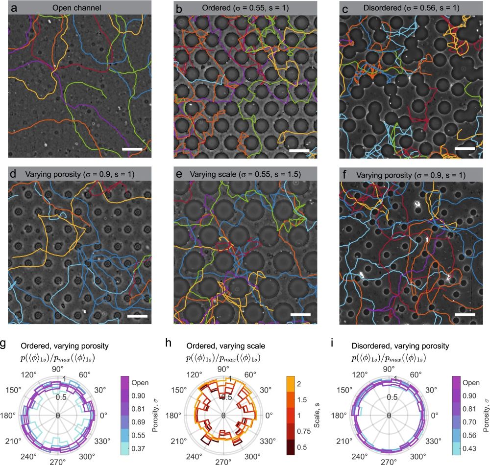
a–f Sample cell trajectories in porous microfluidic devices for various porosity, scale, and disorder of the obstacle lattice. See Table 1 for a full list of porous microfluidic geometries tested. Scale bars, 120 μm. a Cell trajectories in an open channel (porosity, σ = 1). b Ordered porous medium (σ = 0.55) with cylindrical pillars of diameter d = 85 μm, arranged in a hexagonal lattice with a lattice spacing c = 120 μm (scale, s = 1). c Disordered porous medium (σ = 0.55, s = 1), where d and average lattice spacing are similar to (b). d Porosity is varied by modifying the pillar diameters (d = 40 μm), while holding the lattice spacing constant (c = 120 μm). e Identical to (b), but with all features scaled up by a factor s = 1.5. f Identical to (c), but with increased porosity by reducing the pillar diameters to d = 20 μm. g–i Normalized polar probability density of cell swimming directions in various geometries: g ordered lattice with varying porosity (s = 1), h ordered lattice with varying scale (σ = 0.55), and i disordered media with varying porosity (s = 1). Instantaneous swimming direction vectors are averaged over a 1 s interval within the ballistic regime of the cell trajectories to obtain average swimming directions, 〈ϕ〉1s. Cell trajectories were randomly sampled from the entire cell population (Supplementary Fig. 1), and the probability, p(〈ϕ〉1s), is normalized by its maximum value, pmax(〈ϕ〉1s).” Reproduced under Creative Commons Attribution 4.0 International License from Dehkharghani, A., Waisbord, N. & Guasto, J.S. Self-transport of swimming bacteria is impaired by porous microstructure. Commun Phys 6, 18 (2023)..
The research paper focuses on the movement of Magnetococcus marinus bacteria in porous media, with the aim of understanding the mechanisms that regulate self-transport in confined interstices. The researchers employed microfluidic techniques and conducted experiments on microfluidic chips with different geometries.
The results of the study highlight the importance of the effective mean free path in governing cell transport. The researchers found that the reduction of persistence length was the key mechanism hindering transport, and the effective mean free path of the medium was the primary geometric scale that dictated bacterial transport coefficients. The results also show that precise measurements of cell-surface interactions are crucial in understanding long-time transport.
In addition, the researchers developed a theoretical model to predict cell diffusion based on easily measurable parameters, such as cell swimming speed and mean surface scattering angle. The model was consistent with experimental evidence and provides a reliable approach to forecast cell transport properties in porous media.
The study sheds light on how porous microstructure affects the transport of microbes, which is relevant to microbial ecology in sediments and soils. The approach used in the study may also be applied to explore how directed motility affects cell transport through porous microstructures and to investigate a range of micro-swimmers with different surface scattering properties.
The researchers suggest that future studies could extend this work to 3D environments of swimming microbes, using recently developed experimental techniques in microfabrication. The results of the study contribute to a comprehensive understanding of the mechanisms regulating self-transport in porous media and the interactions between cells and surfaces of the solid matrix. The findings of the study provide a foundation for further exploration of the physical ecology of swimming cells and their role in environmental and health hazards in stagnant porous media.
“The effect of directed motility on cell transport through porous microstructures is highly relevant to microbial ecology in sediments and soils, and this approach may provide new insights into magnetotaxis48 and chemotaxis49 in porous media. Although the present experiments and modeling were restricted to quasi-2D microfluidic geometries, recently developed experimental approaches50 could extend this work to model the naturally occurring 3D environments of swimming microbes.“, the authors explained.
Figures and the abstract are reproduced from Dehkharghani, A., Waisbord, N. & Guasto, J.S. Self-transport of swimming bacteria is impaired by porous microstructure. Commun Phys 6, 18 (2023). https://doi.org/10.1038/s42005-023-01136-w
Read the original article: Self-transport of swimming bacteria is impaired by porous microstructure


