17 Feb Microfluidic-based vasculature system for studying tumor-associated properties and drug delivery
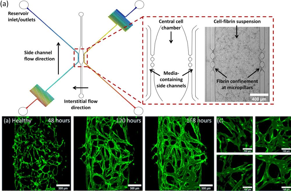
Microfluidic devices have emerged as powerful tools for studying biological processes with high precision and control. In cancer research, the incorporation of a functional vascular system is essential to accurately model the tumor microenvironment and evaluate potential anti-cancer therapeutics. To address this challenge, researchers have developed microfluidic-based vasculature systems that can mimic the properties of tumor-associated blood vessels. These systems are fabricated using microfabrication techniques to create intricate channels and chambers on a microfluidic chip, which can then be seeded with healthy endothelial-fibroblast cell co-cultures and conditioned with tumor cell media to induce the formation of disorganized, tortuous networks with characteristics consistent with those of tumor-associated vasculature. The development of complex 3D tumour cultures has been a major focus in cancer research, as these models provide a more accurate representation of the tumor microenvironment. However, the incorporation of a functional vascular system within these models remains a challenge. The vasculature is crucial in the delivery of therapeutic drugs to the tumor site, and its absence in 3D models limits their ability to accurately predict the efficacy of potential anti-cancer therapies. To address this challenge, in a recent research article published in the Lab on a Chip journal, researchers have utilized a PDMS-based microfluidic vasculature system that can mimic the properties of tumour-associated blood vessels. The system involves conditioning healthy endothelial-fibroblast cell vasculature co-cultures with media taken from tumour cell cultures. This results in the formation of disorganised, tortuous networks that display characteristics consistent with those of tumour-associated vasculature.
“This study reports a simple approach to condition on-chip vasculature to display disease-like characteristics and highlights the importance of producing accurate organ-on-chip models for the successful development of new therapeutics. “, the authors explained.

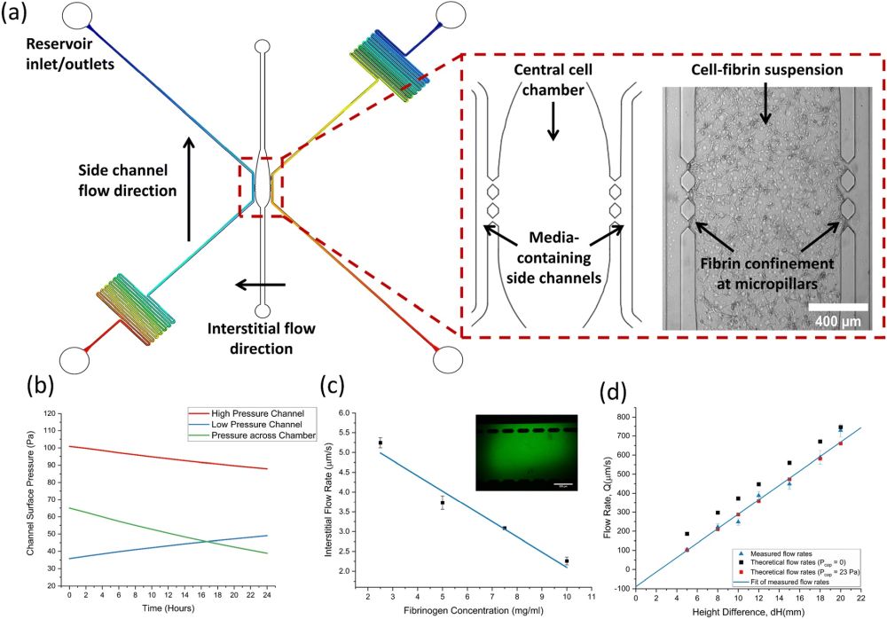
“(a) An AutoCAD outline of the microfluidic device with pressure distribution plots overlayed on the side channels to display pressure equilibration between reservoirs along the channel. The inset shows a close-up of the three-chamber design in AutoCAD, alongside a brightfield image taken after the cell-fibrin suspension was seeded into the central chamber. (b) Graph showing the surface pressure at the pillar gaps in the high- and low-pressure side channels as a function of time. The pressure across the chamber slowly decreases due to decreasing hydrostatic flow. (c) A graph of interstitial flow through empty fibrin gels formed with a range of fibrinogen concentrations. (d) Measured and theoretical flow rates for a range of reservoir height differences. Red data points show theoretical flow rates once the measured capillary pressure value was included in calculations.” Reproduced from M. D. Bourn, S. Z. Mohajerani, G. Mavria, N. Ingram, P. L. Coletta, S. D. Evans and S. A. Peyman, Lab Chip, 2023, Advance Article, DOI: 10.1039/D2LC00963C under Creative Commons Attribution 3.0 Unported Licence.
The researchers found that integrin αvβ3, a cell adhesion receptor associated with angiogenesis, was upregulated in vasculature co-cultures conditioned with tumour cell media (TCM). This is consistent with the reported αvβ3 expression pattern in angiogenic tumour vasculature in vivo. Increased accumulation of liposomes (LSs) conjugated to antibodies against αvβ3 was observed in TCM networks compared to non-conditioned networks, indicating αvβ3 may be a potential target for the delivery of drugs specifically to tumour vasculature. Furthermore, the use of microbubbles (MBs) and ultrasound (US) was investigated to further enhance the delivery of LSs to TCM-conditioned vasculature. The quantification of fluorescent LS accumulation post-perfusion of the vascular network showed a 3-fold increased accumulation with the use of MBs and US, suggesting that targeted LS delivery could be further improved with the use of locally administered MBs and US.
This microfluidic vasculature system provides a promising platform for evaluating potential anti-cancer therapeutics. The characterization of vessel area, diameter, and integrin αvβ3 expression revealed properties consistent with in vivo tumour vasculature. Additionally, the microfluidic system allows for perfusion of model therapeutics exclusively through the vessels, with rates of flow and shear stress similar to those found within capillaries. However, more experiments are still required to further elucidate the efficacy of integrin targeting and MB-mediated drug delivery. Overall, this microfluidic-based vasculature system represents an important step forward in the development of more accurate 3D tumour models and provides a platform for investigating the delivery of anti-cancer therapeutics.
“Whilst the use of indirect tumour conditioning in this study prevented the direct evaluation of tumour cell viability, increased accumulation, and uptake of therapeutics in the vasculature can still be used as an indicator of improved drug delivery. The use of TCM conditioning also enabled the routine production of tumour-associated vasculature compared to previous tumour vasculature systems which have relied on cancer cell-vasculature co-cultures, demonstrating the advantages of this relatively simplistic system. “, the authors explained.
Figures and the abstract are reproduced from M. D. Bourn, S. Z. Mohajerani, G. Mavria, N. Ingram, P. L. Coletta, S. D. Evans and S. A. Peyman, Lab Chip, 2023, Advance Article , DOI: 10.1039/D2LC00963C under Creative Commons Attribution 3.0 Unported Licence
Read the original article: Tumour associated vasculature-on-a-chip for the evaluation of microbubble-mediated delivery of targeted liposomes


