31 Aug Exploring the Stability of Tumor-on-a-Chip Models with Polydopamine Coatings
Pancreatic cancer, notorious for its poor prognosis and rapid progression, remains a significant challenge in oncology. The complexity of pancreatic cancer, particularly pancreatic ductal adenocarcinoma (PDAC), necessitates innovative research approaches to understand and combat this deadly disease. A recent microfluidic chip reported in Biomacromolecules journal aims at the development of stable tumor-on-a-chip models using polydopamine interfacial coating, offering new insights and potential pathways for therapeutic intervention.
“This work proposes using polydopamine (PDA) coating for 3D microfluidic cultures of pancreatic cancer cells to overcome matrix adhesion challenges to sustain representative tumor 3D cultures. Using PDA’s distinctive adhesion and biocompatibility, our model uses type I collagen hydrogels seeded with different pancreatic cancer cell lines, prompting distinct levels of matrix deformation and contraction. “, the authors explained.

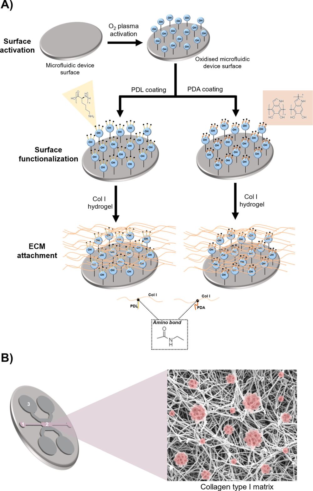
“Experimental set up. (A) Activation and functionalization of PDMS microfluidic devices for long-term 3D culture. Polydimethylsiloxane (PDMS) microfluidic devices were functionalized first by O2 plasma surface activation, followed by coating with either poly-d-lysine (PDL) or polydopamine (PDA). (B) Schematic of the microfluidic device. Single pancreatic cancer cells embedded in a type I collagen hydrogel are introduced through the loading port (1) into the central chamber of the device (2). Through the reservoirs (3), the culture medium is introduced. The isolated cells self-organize three-dimensionally, interacting with the matrix and generating tumor spheroids from single cells.” Reproduced from Soraya Hernández-Hatibi, Pedro Enrique Guerrero, José Manuel García-Aznar, and Elena García-Gareta Biomacromolecules 2024 25 (8), 5169-5180. under a CC BY 4.0 Attribution 4.0 International license
Traditional 2D cell cultures fall short in replicating the three-dimensional structure and complex microenvironment of tumors, which are crucial for accurate biomedical research. Microfluidic devices such as organ-on-a-chips for 3D cultures have emerged as a solution, allowing researchers to mimic the spatial arrangements and interactions of tumor cells more realistically. However, these systems often face challenges with the structural integrity of synthetic extracellular matrices (ECMs) under the mechanical stress exerted by growing tumor cells.
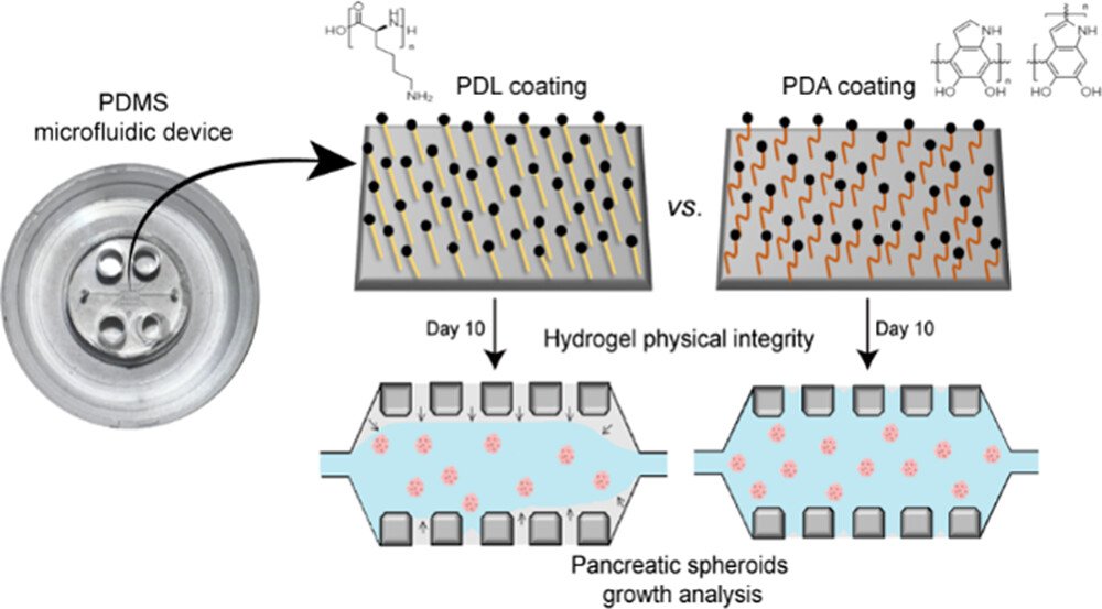
Drawing inspiration from the strong adhesive properties of mussel proteins, the research team employed polydopamine to enhance the adhesion and stability of type I collagen hydrogels within microfluidic devices. Polydopamine facilitates a robust binding that resists the mechanical forces from the tumor cells, thereby maintaining the integrity of the collagen matrix crucial for long-term studies.
The study investigated the optimal concentration and incubation times for PDA coating to achieve a balance that supports both structural stability and the biological activity of tumor cells. It was found that lower concentrations of PDA (0.5 mg/mL) allowed for better cellular dynamics and spheroid development compared to higher concentrations, which could overly restrict cell movements due to stronger adhesion. The study highlighted the use of polydopamine (PDA), a biopolymer inspired by the adhesive proteins of mussels, to enhance the structural integrity of 3D tumor models inside the microfluidic chip. PDA coatings significantly improve the adhesion of collagen hydrogels, crucial for maintaining the integrity of 3D microfluidic cultures. This enhancement allows for extended culture periods without the detachment issues commonly seen with traditional hydrogel-based systems.
Hydrogel Adhesion and Spheroid Integrity:
Adhesion Stability: PDA coatings dramatically enhanced the adhesion of collagen hydrogels, with hydrogels remaining well-confined within the microfluidic device for up to 11 days, unlike those with poly-D-lysine (PDL) which detached due to spheroid-induced contractile forces.
Morphological Consistency: Spheroids in PDA-coated environments maintained more consistent shapes and sizes, showing the ability of PDA to support stable growth dynamics inside the microfluidic platform
Quantitative Growth Dynamics:
Controlled Spheroid Growth: Spheroids in PDA-coated devices showed a steady growth pattern, with an approximate 20% increase in diameter over 10 days, highlighting the platform’s ability to provide a stable environment for tumor growth studies.
By improving the fidelity of 3D tumor models, this approach enhances the potential for accurately studying tumor behavior and testing new cancer treatments. More stable and representative models can lead to better understanding of the interactions between cancer cells and their microenvironment, crucial for the development of targeted therapies. The continued development of such innovative approaches in microfluidics will be critical in bridging the gap between in vitro experiments and real-world clinical outcomes, paving the way for breakthroughs in personalized medicine and effective cancer treatment strategies.
“In summary, the optimization of PDA coatings on polydimethylsiloxane (PDMS) microfluidic devices, as presented in this study, significantly contributes to the advancement of novel in vitro tumor models. These 3D models not only represent a significant advancement in in vitro tumor research but also bridge the gap between traditional 2D cultures and the complex 3D nature of tumors in vivo “, the authors concluded.
Figures are reproduced from Soraya Hernández-Hatibi, Pedro Enrique Guerrero, José Manuel García-Aznar, and Elena García-Gareta Biomacromolecules 2024 25 (8), 5169-5180 DOI: 10.1021/acs.biomac.4c00551 under a CC BY 4.0 Attribution 4.0 International license.
Read the original article: Polydopamine Interfacial Coating for Stable Tumor-on-a-Chip Models: Application for Pancreatic Ductal Adenocarcinoma
For more insights into the world of microfluidics and its burgeoning applications in biomedical research, stay tuned to our blog and explore the limitless possibilities that this technology unfolds. If you need high quality microfluidics chip for your experiments, do not hesitate to contact us.


