11 Feb Exploring the Mechanics of Cancer Cell Metastasis Through Microfluidics
In the fight against cancer, understanding the mechanics of metastasis—the process by which cancer spreads from one part of the body to another—is crucial. Cancer metastasis is a complex process that involves more than just the genetic and biochemical makeup of cancer cells; the physical forces at play also hold significant sway in the progression of the disease. Recent advancements in microfluidics have opened new avenues for exploring this complex phenomenon at the microscopic level. A study published in “Microsystems & Nanoengineering” sheds light on how breast cancer cells navigate the challenging environments of the bloodstream, particularly through the narrow passages of microcapillaries.
“In this study, we first experimentally investigated the deformability of three different breast cancer cell lines (MCF-7, SK-BR-3, and MDA-MB-231) using a constricted microfluidic device whose constriction width is 10 μm. Then, by focusing on the more deformable one (MDA-MB-231), we extended the experiments to two additional constricted microfluidic devices with 8 μm, and 12 μm widths of constriction.“, the authors explained.
The study utilizes microfluidic chips to simulate the physical constraints encountered by circulating tumor cells (CTCs) in microcapillaries. These microfluidic devices, engineered through microfluidic microfabrication techniques, provide a controlled setting for observing the behavior of cancer cells under various flow conditions. The precision and scalability of microfluidics make it an indispensable tool in these research, offering insights into the physical forces at play during the metastasis process.
A key finding from the research is the variation in cancer cell deformability, particularly how it relates to metastatic potential. The experiments conducted using the PDMS-based microfabricated microfluidic chips reveal that cells with higher metastatic capabilities are more deformable, allowing them to navigate through tighter spaces more effectively. This observation is critical, as it suggests that the physical properties of cancer cells could be indicative of their ability to metastasize, providing a potential biomarker for assessing cancer aggressiveness.

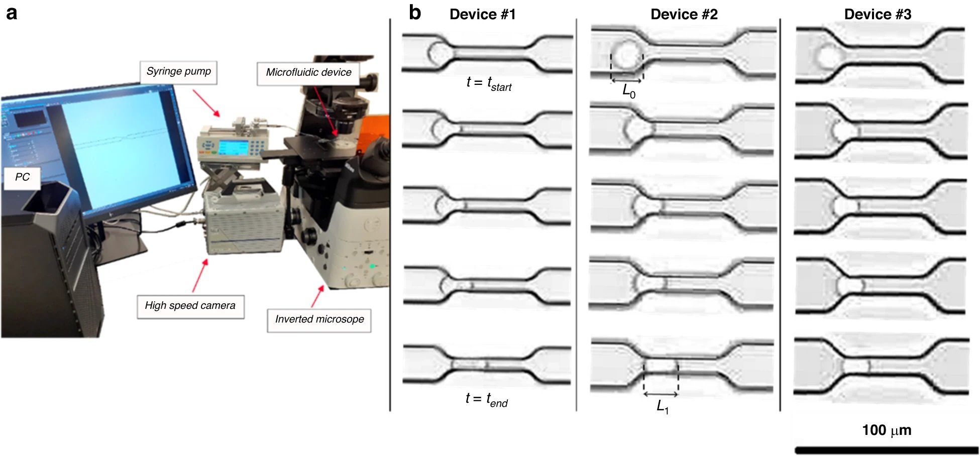
“a Experimental set up for high-speed measurements of the single breast cancer cell deformability. b Captured images from entry process of the single breast cancer cell with the diameter of 18.2 µm (passing device #1), 22.3 µm (passing device #2) and 17.9 µm (passing device #3) at five instances using the three constricted microfluidic devices ” Reproduced from Keshavarz Motamed, P., Abouali, H., Poudineh, M. et al. Experimental measurement and numerical modeling of deformation behavior of breast cancer cells passing through constricted microfluidic channels. Microsyst Nanoeng 10, 7 (2024). https://doi.org/10.1038/s41378-023-00644-7 under a CC BY 4.0 DEED Attribution 4.0 International license.
To complement the experimental observations, the study employs numerical modeling techniques to simulate the deformation behavior of cancer cells within the microfluidic channels. This approach allows for a deeper understanding of the cellular mechanics involved and offers predictive insights that could inform the development of new therapeutic strategies. The integration of experimental data with computational models underscores the power of interdisciplinary research in tackling complex biological questions.
The implications of this study extend beyond the immediate findings. By demonstrating the capability of microfluidic devices to replicate and study the microenvironments encountered by cancer cells, it paves the way for further explorations into the mechanical aspects of cancer metastasis. The use of microfluidics in cancer research promises to enhance our understanding of disease progression, improve diagnostic methods, and lead to the development of more targeted and effective treatments.

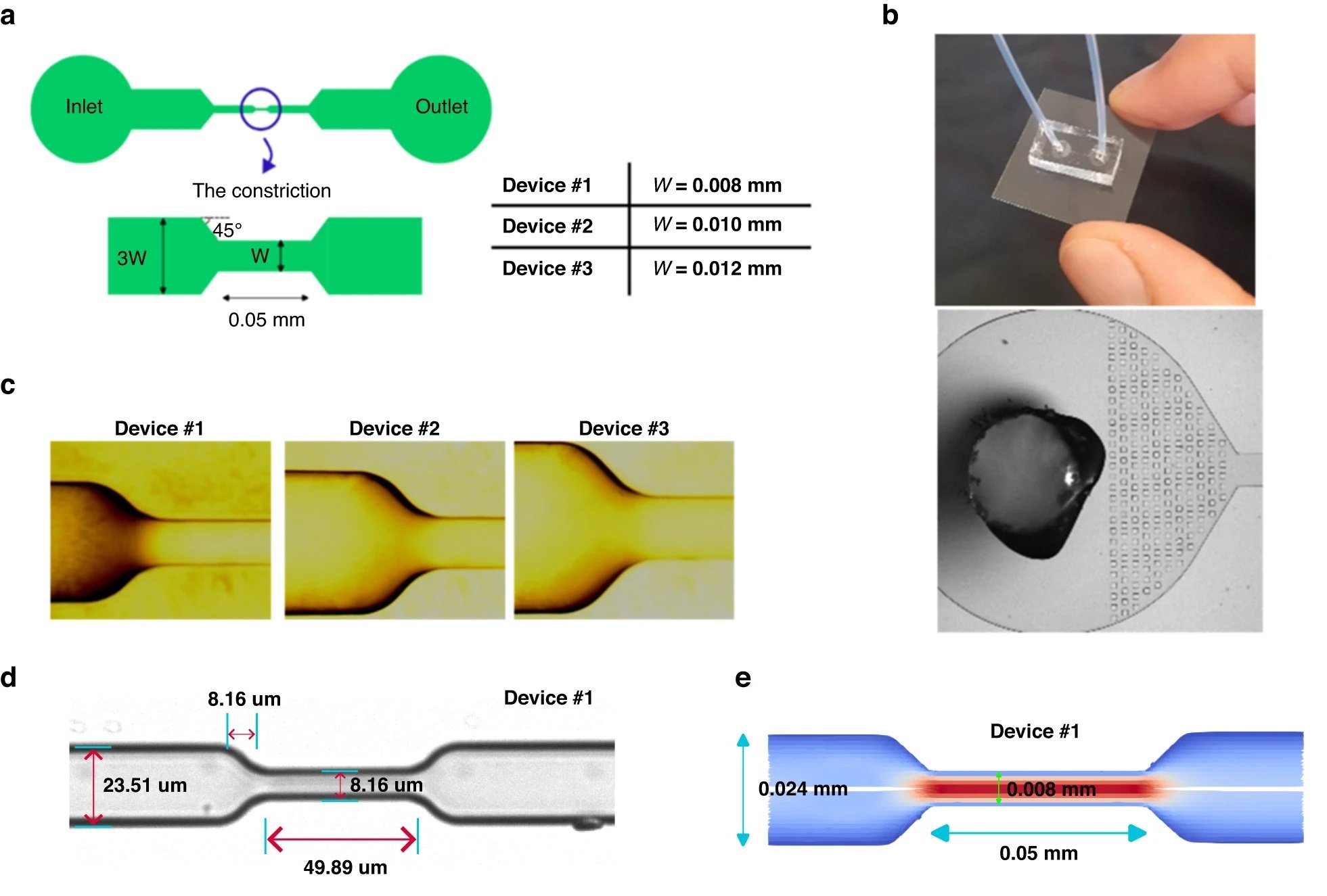
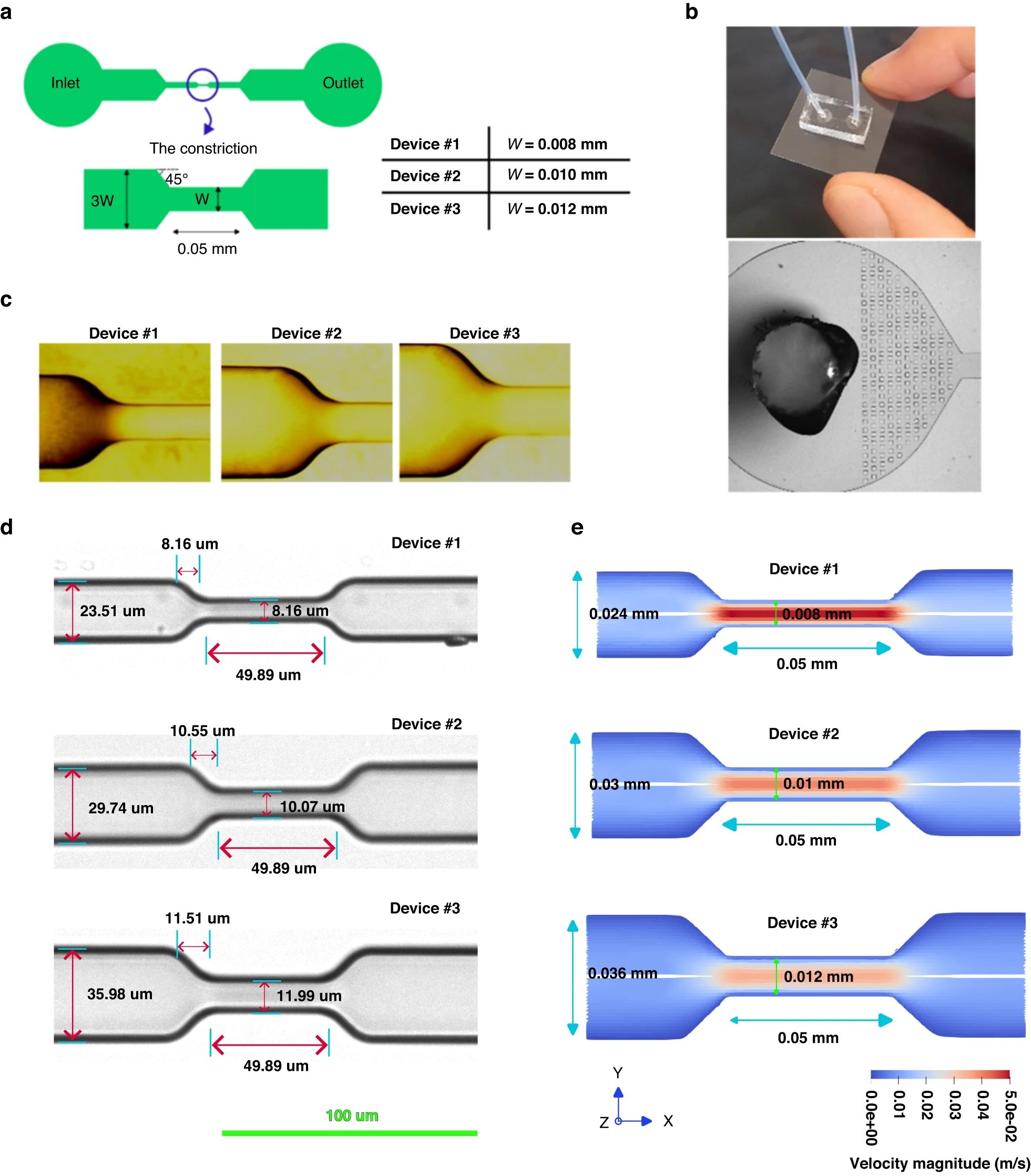
“a Schematic representation of the microfluidic devices design. b Actual image of the designed one-channel device and the filters devised in the inlet. c Magnified view of the entrance section of the constricted channels in the fabricated mold using 100× objective. d Magnified view of the constricted channel in the fabricated devices using 20× objective. e The fluid velocity magnitude at the mid-plane in the Z direction for 20 μL/h flow rate extracted from 3D CFD simulations of each constricted channel” Reproduced from Keshavarz Motamed, P., Abouali, H., Poudineh, M. et al. Experimental measurement and numerical modeling of deformation behavior of breast cancer cells passing through constricted microfluidic channels. Microsyst Nanoeng 10, 7 (2024). https://doi.org/10.1038/s41378-023-00644-7 under a CC BY 4.0 DEED Attribution 4.0 International license.
This study represents a step forward in our understanding of cancer metastasis. By leveraging the capabilities of microfluidic technology, researchers can now explore the physical dynamics of cancer cell movement with unprecedented detail and precision. As we continue to explore the interface between engineering and biology, the hope for more effective cancer diagnostics and therapies becomes increasingly tangible.
“Since the mechanical properties of each cancer cell type is different from other cancer cell types, the combined experimental-numerical method, proposed in this work, can be used to obtain the valid model specific to that cell type. Such models can be used to investigate the situation in which cancer cells physically occlude a microcapillary or adhere to a vessel wall can be studied by applying the validated model presented here. Furthermore, the motion and deformation behavior of CTC clusters can be numerically obtained by repeating the presented approach for CTC clusters. “, the authors concluded.
Figures are reproduced from Keshavarz Motamed, P., Abouali, H., Poudineh, M. et al. Experimental measurement and numerical modeling of deformation behavior of breast cancer cells passing through constricted microfluidic channels. Microsyst Nanoeng 10, 7 (2024). https://doi.org/10.1038/s41378-023-00644-7 under a CC BY 4.0 DEED Attribution 4.0 International license.
Read the original article: Experimental measurement and numerical modeling of deformation behavior of breast cancer cells passing through constricted microfluidic channels
For more insights into the world of microfluidics and its burgeoning applications in biomedical research, stay tuned to our blog and explore the limitless possibilities that this technology unfolds. If you need high quality microfluidics chip for your experiments, do not hesitate to contact us.


