01 Sep Droplet Microfluidics with Matrix-Assisted Laser Desorption/Ionization-Mass Spectrometry (MALDI)-MSDetection
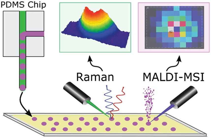
“Microfluidic and mass spectrometry (MS) methods are widely used to sample and probe the chemical composition of biological systems to elucidate chemical correlates of their healthy and disease states. Though matrix-assisted laser desorption/ionization-mass spectrometry (MALDI)-MS has been hyphenated to droplet microfluidics for offline analyses, the effects of parameters related to droplet generation, such as the type of oil phase used, have been understudied. To characterize these effects, five different oil phases were tested in droplet microfluidics for producing samples for MALDI-MS analysis. Picoliter to nanoliter aqueous droplets containing 0.1 to 100 mM γ-aminobutyric acid (GABA) and inorganic salts were generated inside a polydimethylsiloxane microfluidic chip and deposited onto a conductive glass slide. Optical microscopy, Raman spectroscopy, and MALDI-mass spectrometry imaging (MSI) of the droplet samples and surrounding areas revealed patterns of solvent and oil evaporation and analyte deposition. Optical microscopy detected the presence of salt crystals in 50–100 μm diameter dried droplets, and Raman and MSI were used to correlate GABA signals to the visible droplet footprints. MALDI-MS analyses revealed that droplets prepared in the presence of octanol oil led to the poorest detectability of GABA, whereas the oil phases containing FC-40 provided the best detectability; GABA signal was localized to the footprint of 65 pL droplets with a limit of detection of 23 amol. The effect of the surfactant perfluorooctanol on analyte detection was also investigated..”

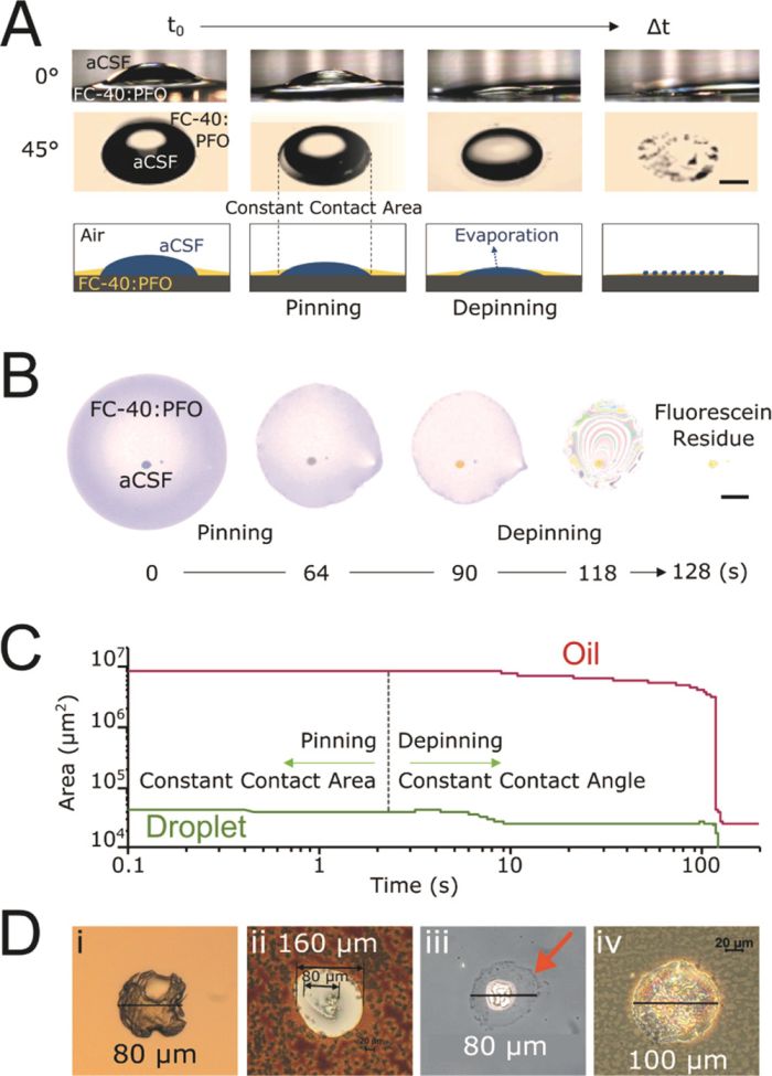
“Figure 1. Dynamics of droplet drying and morphology on an ITO glass slide. Evaporation of picoliter sessile droplets and surrounding oil are responsible for size reduction of visible structures. (A) Time-lapsed microscope images of 500 nL aqueous droplet and FC40:PFO oil phase at a 0° and 45° angle projections. Scale bar, 500 μm. (B) Time-lapsed images of evaporation of 65 pL droplet containing 100 μM fluorescein and 100 μM GABA surrounded by oil phase. Scale bar, 150 μm. (C) Time course of pinning and depinning phases of droplet on ITO substrate. Droplet and oil are completely dried in 10 and 100 s, respectively. (D) Dried cores of droplets produced in FC-40:PFO for 1000 pL volumes (i) before matrix and (ii) after matrix application; and for 65 pL volumes (iii) before matrix and (iv) after matrix. The oil residue halo surrounding the droplet core is visible in (iii) (marked by an orange arrow).” Reproduced under Creative Commons Attribution 4.0 International License. Bell et al., ACS Meas. Sci. Au., 2021.
Read the original article: Droplet Microfluidics with MALDI-MS Detection: The Effects of Oil Phases in GABA Analysis


