04 Feb Microfluidic Research on C. elegans for Developmental Toxicity Testing: A Novel Machine Learning Approach
Traditional developmental toxicity (DevTox) studies largely rely on mammalian models to assess chemical impacts on development. These methods, while comprehensive, require extensive resources and time. There is a growing need for more efficient alternatives to reduce reliance on vertebrate models and enhance throughput capabilities in toxicological assessments. The introduction of a microfluidic chip called vivoChip marks an advancement in developmental toxicity testing. This microfluidic device can immobilize and process large numbers of C. elegans rapidly, providing a more efficient and less resource-intensive solution. The vivoChip allows for the collection of high-resolution, 3D images of approximately 1000 C. elegans across 24 populations, facilitating detailed analysis without the need for anesthetics, which can affect measurement accuracy.
“With the recent advent of the large-scale microfluidic device, vivoChip, we can now rapidly collect 3D high-resolution images of ~ 1000 C. elegans from 24 different populations. “, the authors explained.
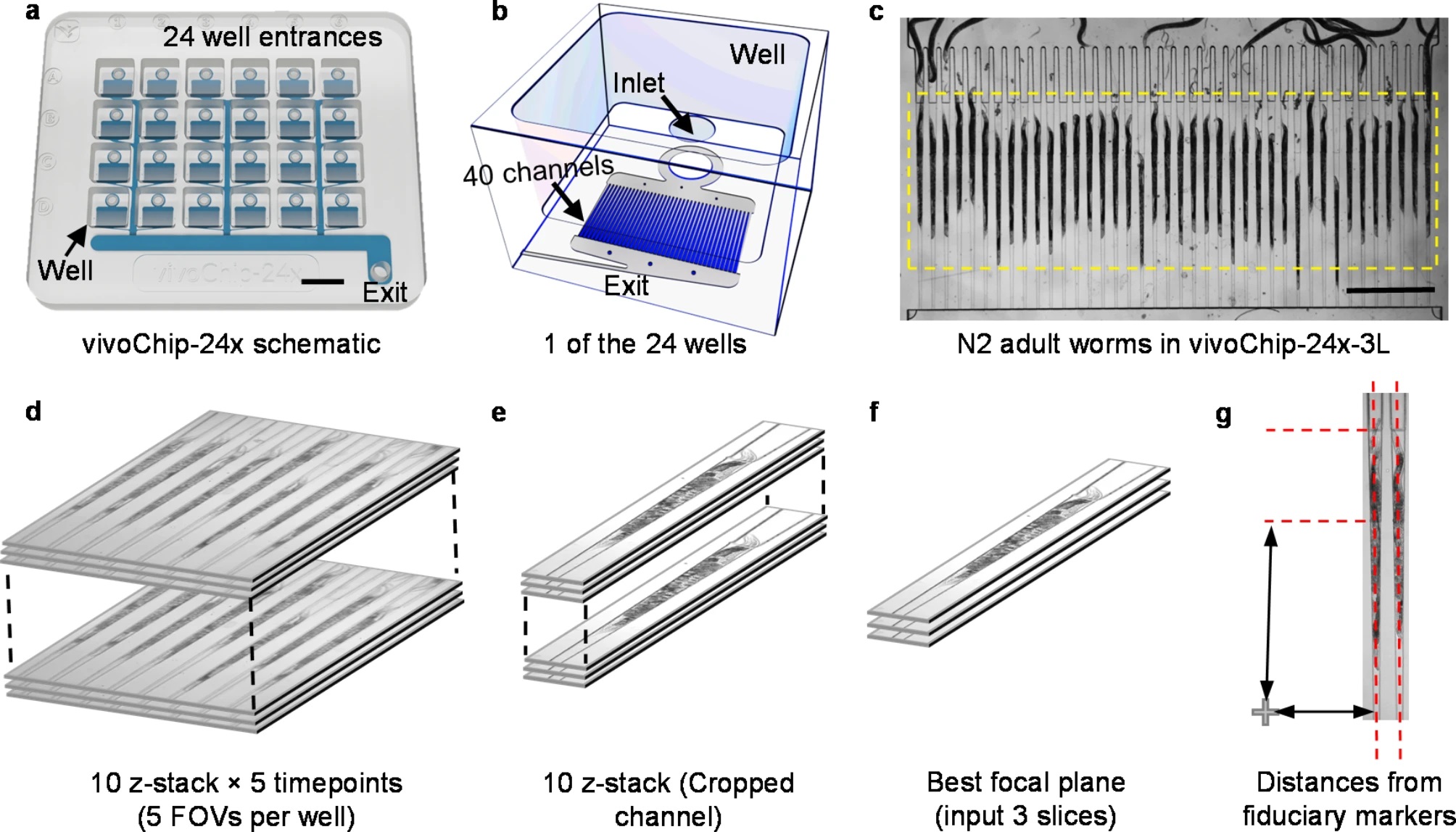
“High-resolution C. elegans body images are acquired using a vivoChip-24x platform. (a) Schematic of vivoChip-24x technology designed to immobilize C. elegans and capture high-resolution images from 24 different populations with up to 40 worms per population. Scale bar = 9 mm. (b) Detailed schematic of a single well from the 24-well device. Each well features an opening at the top and an inlet on the bottom, leading to 40 parallel immobilization channels beneath it. Worms are loaded into the well, flow through the inlet, and are immobilized within the 40 channels by the pressure of the fluid flow directed toward the exit. (c) Brightfield image of 40 adult C. elegans immobilized inside the microfluidic channels of the vivoChip-24x-3L device. Scale bar = 1 mm. (d) Graphic demonstrating a z-stack of 10 images collected for each FOV. Each FOV covers 8 channels using a 10 × , 0.4 NA objective. For each FOV, we collect a total of 50 images over 5 time points, with 10 z-slices (6 µm z-step size) captured at each time point (1-s interval). (e) We crop each channel for the first time point. (f) We identify the best focal plane for analysis and use 3 slices, comprising the focal plane, one slice above, and one slice below to train the network. (g) The location of the worm within the channel is used to determine the channel height by referencing its position with respect to the fiduciary cross mark present within each well of the vivoChip-24x.” Reproduced from DuPlissis, A., Medewar, A., Hegarty, E. et al. Machine learning-based analysis of microfluidic device immobilized C. elegans for automated developmental toxicity testing. Sci Rep 15, 15 (2025). under a CC BY 4.0 Attribution 4.0 International license
The vivoChip efficiently immobilizes C. elegans in a microfluidic setup that enables parallel processing of multiple samples. This system captures detailed morphological data of C. elegans, which are crucial for high-resolution developmental toxicity testing. The introduction of a machine-learning platform, vivoBodySeg, employs a 2.5D U-Net architecture to automate image segmentation. This method significantly reduces the time required for image analysis, processing 36 GB of data per device in about 35 minutes, which is about 140 times faster than manual processing.
The machine learning model achieves a high Dice score of 97.80%, indicating excellent accuracy in segmenting C. elegans from the images. This model not only expedites the process but also enhances reproducibility and precision in measuring developmental toxicity parameters.
The integration of microfluidic technology with advanced machine learning algorithms offers a transformative approach to developmental toxicity testing. This method significantly reduces reliance on traditional mammalian models, lowers resource usage, and increases throughput, all while maintaining high accuracy and reproducibility. The vivoChip and the associated vivoBodySeg platform represent significant steps forward in the field of toxicological research, promising faster and more ethical approaches to chemical safety evaluation.
“Our ML pipeline, combined with scalable microfluidic technology, provides a rapid, high-throughput solution for DevTox studies, offering automated, precise quantifications of C. elegans volume, which is analogous to body weight assessments in mammalian tests. In the future, we will test a large number of reference chemicals and estimate the accuracy and sensitivity by comparing them with other species “, the authors concluded
Figures are reproduced from Mishra, A., Huang, SB., Dubash, T. et al. Tumor cell-based liquid biopsy using high-throughput microfluidic enrichment of entire leukapheresis product. Nat Commun 16, 32 (2025). https://doi.org/10.1038/s41467-024-55140-x under a CC BY 4.0 Attribution 4.0 International license.
Read the original article: Machine learning-based analysis of microfluidic device immobilized C. elegans for automated developmental toxicity testing
For more insights into the world of microfluidics and its burgeoning applications in biomedical research, stay tuned to our blog and explore the limitless possibilities that this technology unfolds. If you need high quality microfluidics chip for your experiments, do not hesitate to contact us.