06 Feb A microphysiological system that can benefit oncolytic virus therapy by mimicking the bystander effect
A major challenge in cancer therapy is tumor-targeted drug delivery. Targeted drug delivery refers to the accumulation of a drug near a specific zone to maximize the therapeutic effect. Over the years various strategies have been developed to increase the efficiency of the targeted drug delivery to solid tumors and reduce drug-related toxicity. Oncolytic virus (OV) therapy is one of these methods that hinges on employing natural or synthetic viruses that preferentially attack cancer cells. A better understanding of the events involved in the transport of the drug after injection as well as what occurs after the drug being delivered is essential for characterizing cancer therapy strategies such as oncolytic virus therapy.
When a cancer cell gets infected by a replicating OV, the virus starts making copies until the cell bursts. The bursting of the cell causes copies of the virus along with cancer-specific antigens to be released. The released viruses or virions can infect other surrounding cancer cells to destroy the remaining cancer cells. The antigens can also alert the immune system giving rise to a local response. However, the OVs do more than just destroying the cancer cells. They can also affect the uninfected cells in the vicinity and cause apoptosis. This phenomenon is called the bystander effect.
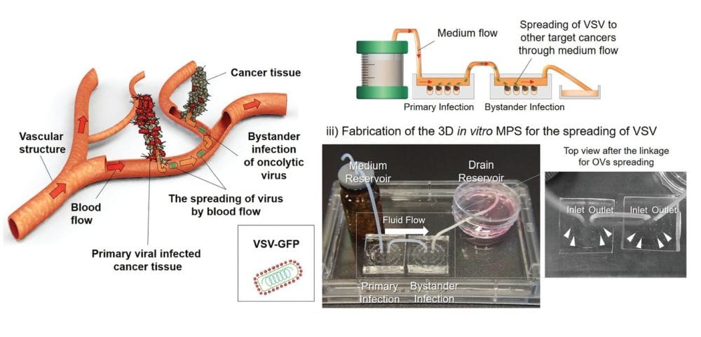
A microphysiological system (MPS) capable of mimicking the in vivo events such as the bystander effect can help in devising novel oncoselective strategies for more effective tumor destruction. Researchers now have developed a platform that gives a better insight into oncolytic viruses. In a research article published in the Advanced Biomaterials journal, a group of researchers reported a microfluidic device for evaluating the bystander effect caused by oncolytic viruses.
The reported microfluidics platform included two microfluidic chips connected with Teflon tubing. In the first chip, they created a multicellular tumoroid (MCT) environment that includes human lung cancer cells, human lung fibroblasts, human umbilical vein endothelial cells, and ECM. The primary MPS was exposed to vesicular stomatitis virus (VSV) tagged with a green fluorescent protein (GFP) for one day. Next, the second MPS was connected to the primary device to observe the bystander effect in the second MPS. By connecting the two chips and transferring the medium from the first chip to the second, they could mimic the bystander phenomenon and analyze its effects on the cells in the second chip.
In their conclusion remark, authors mentioned: “We expect that this 3D in vitro MPS will be useful for studying oncotarget delivery using various types of OVs and cancers as a system that can evaluate spreading and immune-related responses induced by viral infection.”
Read the original research article: Evaluation of Bystander Infection of Oncolytic Virus using a Medium Flow Integrated 3D In Vitro Microphysiological System

Pouriya Bayat
Pouriya is a microfluidic production engineer at uFluidix. He received his B.Sc. and M.A.Sc. both in Mechanical Engineering from Isfahan University of Technology and York University, respectively. During his master's studies, he had the chance to learn the foundations of microfluidic technology at ACUTE Lab where he focused on designing microfluidic platforms for cell washing and isolation. Upon graduation, he joined uFluidix to even further enjoy designing, manufacturing, and experimenting with microfluidic chips. In his free time, you might find him reading a psychology/philosophy/fantasy book while refilling his coffee every half an hour. Is there a must-read book in your mind, do not hesitate to hit him up with your to-read list.


